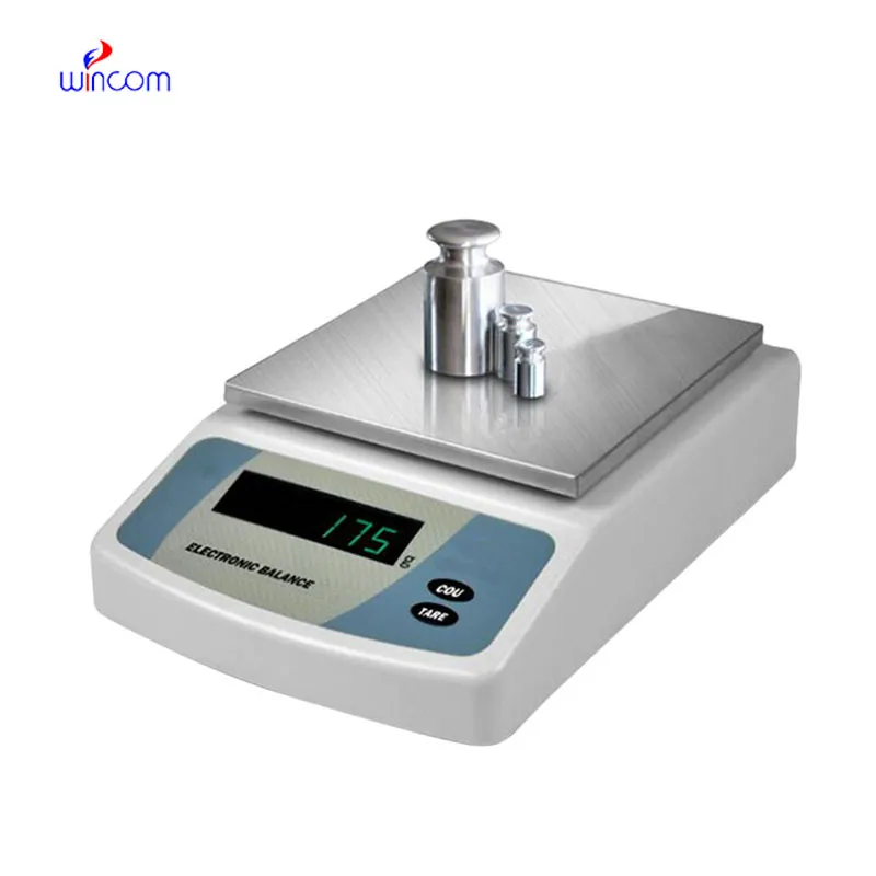
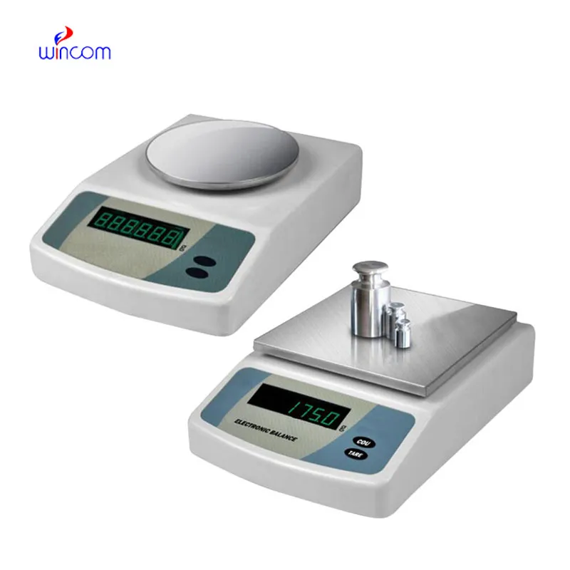
Electronic Balance ensures accurate weighing of clinical sample preparation, research studies, and hospital and laboratory medication formulation. It has an application envisaged to measure even the tiniest amounts with excellent sensitivity and repeatability. Electronic Balance is the instrument laboratory technicians trust for maintaining the highest level of accuracy in analysis, validation of experimental practices, and patients' care support. The use of this instrument in the lab operations not only guarantees results that are reliable but also creates a consistent workflow and quality control which is improved in both the diagnostic and research environments.

Electronic Balance is commonly used in the compounding of medicinal and chemical substances in small quantities in hospital pharmacy laboratories. Mass control with high precision is critical when dealing with active pharmaceutical ingredients in micro or milligram quantities. This application helps to prepare exact doses, internal validation, and experimental drug research. By allowing for repeatable measurements, Electronic Balance helps pharmacists and researchers in controlling formulation processes, which makes it easier to get consistency and reliability throughout hospital medication development workflows.
Electronic Balance will envision a new era where the user-centered interfaces of hospital workers take greater priority. Such friendly visualizations, accompanied by the directing of workflows and notifications, will prove to be helpful during the working of professionals with machines. This change will cut down the duration of training and will also bring in better quality of work in clinical laboratories. Electronic Balance will not give up on the accuracy aspect while still allowing the ease of use in the hospital environments that are changing fast.
For Electronic Balance to last long, professionals must do scheduled servicing as per the laboratory guidelines. Internal cleaning and checking of parts will help to slow down and eventually stop the performance degradation. Hospitals or labs that maintain structured service intervals not only enjoy lesser downtimes but also higher accuracy, thus they can carry on with seamless analytical operations.
Electronic Balance plays an important role in the hospital pharmacy in the accurate formulation of medications, intravenous solutions, and compounded drugs. Even slight alterations in the weight of drugs can change the effectiveness of the drug and endanger the patient's safety. The Pharmacy Technicians rely on Electronic Balance for the correct dosing and checking of the active ingredients. The tool's accuracy guarantees the dependable preparation of drugs, the observance of the rules, and the quality control of the overall hospital pharmacy activities.
Q: What maintenance does an Analytical Balance require? A: A periodic cleaning, checking of the calibration, and also verifying the performance are all necessary. Q: Can an Analytical Balance handle continuous daily use? A: Yes, provided that the correct laboratory conditions and rules are followed. Q: Why is leveling important for an Analytical Balance? A: The accuracy and repeatability of the measurements depend on proper leveling. Q: Can Analytical Balances be connected to laboratory systems? A: Most of the models allow connectivity with laboratory information systems. Q: Are Analytical Balances sensitive to vibration? A: Yes, stable weight readings can be disturbed by vibrations.
We’ve used this centrifuge for several months now, and it has performed consistently well. The speed control and balance are excellent.
This ultrasound scanner has truly improved our workflow. The image resolution and portability make it a great addition to our clinic.
To protect the privacy of our buyers, only public service email domains like Gmail, Yahoo, and MSN will be displayed. Additionally, only a limited portion of the inquiry content will be shown.
We’re interested in your delivery bed for our maternity department. Please send detailed specifica...
Could you share the specifications and price for your hospital bed models? We’re looking for adjus...
E-mail: [email protected]
Tel: +86-731-84176622
+86-731-84136655
Address: Rm.1507,Xinsancheng Plaza. No.58, Renmin Road(E),Changsha,Hunan,China

 af
af
 es
es
 ar
ar
 tr
tr
 sw
sw
 pt
pt
 th
th
 ur
ur
 bn
bn
 ne
ne
 vi
vi
 km
km
 lo
lo
 de
de
 ru
ru
 fi
fi
 nl
nl
 fa
fa
 fr
fr
 ko
ko