With the cutting-edge imaging processors, the doppler fetal doppler facilitates real-time, high-resolution images that are paramount in the detection of subtle physiological changes by clinicians. The display is user-friendly for easy parameter modification as well as image marking. The doppler fetal doppler exhibits the mix of effectiveness, mobility, and reliability for a huge range of diagnostic procedures.

In emergency departments, the doppler fetal doppler is used for instant imaging to easily spot internal wounds and bleeding. It supports the doctor with the abdominal trauma and chest condition diagnosis. Moreover, the doppler fetal doppler provides assistance in rural and field medical practice, delivering consistent imaging in areas with poor medical facilities.

The doppler fetal doppler will proceed to develop as new innovations emerge in artificial intelligence and data analysis. The new models of the doppler fetal doppler will be able to provide training simulations that experts can use to improve scanning sessions. The increased processing power and connectivity of the doppler fetal doppler will set new standards of accessibility and accuracy in medical scanning.

In order to retain the accuracy of the doppler fetal doppler, it is important for operators to check the cables and connections of the transducers for evidence of wear. After each use, the surfaces should be wiped clean using non-abrasive cleaners. The doppler fetal doppler should be turned off properly and covered to prevent dust from collecting. Regular checks by trained personnel should be done.
Used in hospitals and clinics, the doppler fetal doppler provides immediate visual feedback for a variety of medical evaluation uses. Converting sound waves into live images, the doppler fetal doppler allows physicians to easily detect abnormalities. The doppler fetal doppler assists with making diagnostic processes safer in addition to improving patient outcomes. It possesses an ergonomic shape alongside digital integration capabilities that support simple data sharing and medical record documentation.
Q: What imaging modes are available on the ultrasound scannert? A: It supports multiple modes such as B-mode, M-mode, and color Doppler for diverse diagnostic applications. Q: How does the ultrasound scannert improve diagnostic accuracy? A: By providing high-resolution images and real-time feedback, it enables more precise medical evaluations. Q: Can the ultrasound scannert be used in field or remote settings? A: Yes, its portable versions are designed for mobility and can be used in clinics, hospitals, or mobile healthcare units. Q: What kind of display does the ultrasound scannert use? A: It typically features a high-definition digital display that enhances image visualization and readability. Q: How is data from the ultrasound scannert managed? A: The device allows secure storage, easy access, and export of imaging data through USB or network connections.
I’ve used several microscopes before, but this one stands out for its sturdy design and smooth magnification control.
The water bath performs consistently and maintains a stable temperature even during long experiments. It’s reliable and easy to operate.
To protect the privacy of our buyers, only public service email domains like Gmail, Yahoo, and MSN will be displayed. Additionally, only a limited portion of the inquiry content will be shown.
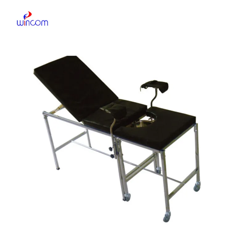
Could you share the specifications and price for your hospital bed models? We’re looking for adjus...
Hello, I’m interested in your water bath for laboratory applications. Can you confirm the temperat...
E-mail: [email protected]
Tel: +86-731-84176622
+86-731-84136655
Address: Rm.1507,Xinsancheng Plaza. No.58, Renmin Road(E),Changsha,Hunan,China

 af
af
 es
es
 ar
ar
 tr
tr
 sw
sw
 pt
pt
 th
th
 ur
ur
 bn
bn
 ne
ne
 vi
vi
 km
km
 lo
lo
 de
de
 ru
ru
 fi
fi
 nl
nl
 fa
fa
 fr
fr
 ko
ko