The fetal doppler 12 weeks combines state-of-the-art digital imaging algorithms that boost contrast and depth perception, which leads to soft tissues being visualized more clearly. The intelligent interface features customizable scanning modes and user profiles for easier operation. Due to its low power consumption and sturdy construction, the fetal doppler 12 weeks maintains performance that is even in high-demand clinical environments.

The fetal doppler 12 weeks has a very wide use in radiology where it supports the guidance of non-invasive procedures. It is very important in gynecology where it is allowed to conduct reproductive system evaluations. In orthopedics, the fetal doppler 12 weeks helps visualize muscles, joints, and tendons ensuring correct diagnostic interpretation. It is this very nature of versatility that makes it a must-have for imaging during procedures in real time.

The fetal doppler 12 weeks should integrate with intelligent diagnostic ecosystems and communicate effortlessly with smartphones and electronic records. The synchronized exchange of data in real-time should enable constant patient observation. The next version should focus on improved design, better processing power of artificial intelligence algorithms, and enhanced reconstruction functions.

Proper upkeep of the fetal doppler 12 weeks helps maintain both the safety of the users as well as the durability of the equipment. The equipment's ventilation and power components must also be regularly inspected for evidence of obstruction and wear. In order to maintain the continued high-quality output of images from the fetal doppler 12 weeks, it must be properly maintained.
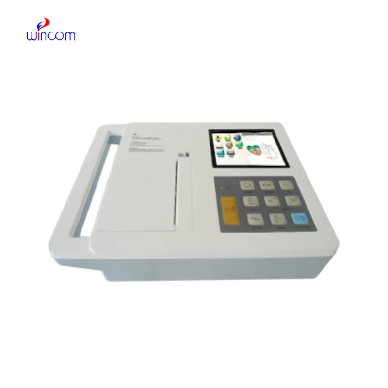
The fetal doppler 12 weeks uses state-of-the-art ultrasound technology to deliver real-time imaging for diagnostic and monitoring purposes. It aids physicians in assessing organs, blood vessels, and soft tissue with unmatched clarity. The non-surgical device is an important tool for guiding medical procedures and making precise diagnoses. The fetal doppler 12 weeks combines portability with precision, rendering it extremely useful in routine exams as well as emergency applications.
Q: How does the ultrasound scannert contribute to emergency diagnostics? A: It enables rapid assessment of internal injuries and organ conditions in time-sensitive situations. Q: Can the ultrasound scannert be upgraded with new features? A: Yes, most models support software updates to enhance performance and expand diagnostic functions. Q: What kind of power supply does the ultrasound scannert use? A: It operates on standard AC power and may include rechargeable battery options for mobile use. Q: Is the ultrasound scannert compatible with electronic medical record systems? A: Yes, it can connect to EMR systems to streamline patient data entry and storage. Q: What factors influence the image quality of the ultrasound scannert? A: Image quality depends on probe type, operator technique, and the frequency settings selected for scanning.
The centrifuge operates quietly and efficiently. It’s compact but surprisingly powerful, making it perfect for daily lab use.
This x-ray machine is reliable and easy to operate. Our technicians appreciate how quickly it processes scans, saving valuable time during busy patient hours.
To protect the privacy of our buyers, only public service email domains like Gmail, Yahoo, and MSN will be displayed. Additionally, only a limited portion of the inquiry content will be shown.
Could you share the specifications and price for your hospital bed models? We’re looking for adjus...
We’re currently sourcing an ultrasound scanner for hospital use. Please send product specification...
E-mail: [email protected]
Tel: +86-731-84176622
+86-731-84136655
Address: Rm.1507,Xinsancheng Plaza. No.58, Renmin Road(E),Changsha,Hunan,China

 af
af
 es
es
 ar
ar
 tr
tr
 sw
sw
 pt
pt
 th
th
 ur
ur
 bn
bn
 ne
ne
 vi
vi
 km
km
 lo
lo
 de
de
 ru
ru
 fi
fi
 nl
nl
 fa
fa
 fr
fr
 ko
ko