The fetal heart doppler combines state-of-the-art digital imaging algorithms that boost contrast and depth perception, which leads to soft tissues being visualized more clearly. The intelligent interface features customizable scanning modes and user profiles for easier operation. Due to its low power consumption and sturdy construction, the fetal heart doppler maintains performance that is even in high-demand clinical environments.

The vast clinical applications of the fetal heart doppler technology made it possible for nephrology to monitor kidney function efficiently and detect abnormalities in kidney structure. In the frontiers of endocrinology, the obtained data can reveal even the smallest nodules in the glands. The fetal heart doppler is also a surgical device for blood flow patterns and vessel integrity.

The fetal heart doppler should integrate with intelligent diagnostic ecosystems and communicate effortlessly with smartphones and electronic records. The synchronized exchange of data in real-time should enable constant patient observation. The next version should focus on improved design, better processing power of artificial intelligence algorithms, and enhanced reconstruction functions.

Care of the fetal heart doppler involves much more than cleanup. Environmental monitoring and mechanical protection are also part of the process. The fetal heart doppler should not be subject to either vibration or shock. The fetal heart doppler should be backed up periodically to retain vital images.
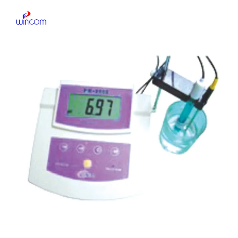
The fetal heart doppler is an essential diagnostic modality in the modern healthcare system, permitting the non-invasive imaging of internal organs and tissues. By transmitting sound waves and reading their echoes, it provides real-time data on physiology. The fetal heart doppler makes precise diagnoses feasible in all specialties, improving clinical decision-making and patient confidence.
Q: What are the main maintenance requirements for the ultrasound scannert? A: Regular cleaning, proper probe handling, and scheduled inspections help maintain optimal performance. Q: How often should the ultrasound scannert be calibrated? A: Calibration frequency depends on usage levels, but periodic professional checks are recommended. Q: Is the ultrasound scannert suitable for pediatric use? A: Yes, it provides gentle, non-invasive imaging ideal for neonatal and pediatric diagnostics. Q: Does the ultrasound scannert support wireless connectivity? A: Many models include Wi-Fi or Bluetooth features for data sharing and device integration. Q: What materials are used in the ultrasound scannert construction? A: It is built with durable medical-grade components designed to withstand continuous clinical use.
The hospital bed is well-designed and very practical. Patients find it comfortable, and nurses appreciate how simple it is to operate.
The centrifuge operates quietly and efficiently. It’s compact but surprisingly powerful, making it perfect for daily lab use.
To protect the privacy of our buyers, only public service email domains like Gmail, Yahoo, and MSN will be displayed. Additionally, only a limited portion of the inquiry content will be shown.
We’re interested in your delivery bed for our maternity department. Please send detailed specifica...
We are planning to upgrade our imaging department and would like more information on your mri machin...
E-mail: [email protected]
Tel: +86-731-84176622
+86-731-84136655
Address: Rm.1507,Xinsancheng Plaza. No.58, Renmin Road(E),Changsha,Hunan,China

 af
af
 es
es
 ar
ar
 tr
tr
 sw
sw
 pt
pt
 th
th
 ur
ur
 bn
bn
 ne
ne
 vi
vi
 km
km
 lo
lo
 de
de
 ru
ru
 fi
fi
 nl
nl
 fa
fa
 fr
fr
 ko
ko