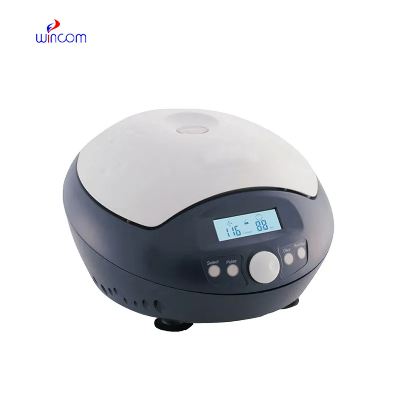
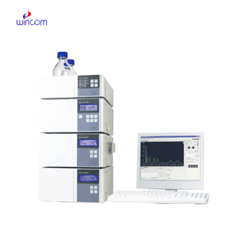
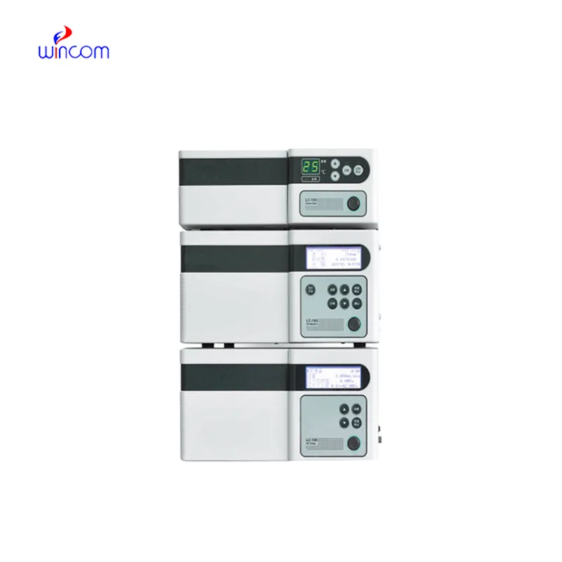
hplc laboratory is a primary tool in hospital and laboratory analytics. Its skills of isolating, measuring, and characterizing both chemical and biological substances enhance research as well as clinical testing. Quality control, drug testing, and testing of samples are done by laboratory technicians using hplc laboratory. The device's flexibility and reliability guarantee uniform performance, yielding critical analytical data that are vital for patient care, experimental validation, and smooth and fast laboratory operations in both healthcare and scientific domains.

Hospital laboratories depend on hplc laboratory for identifying minute quantities of pharmaceuticals and therapeutic agents in difficult-to-analyze biological samples. Its use spans drug compliance testing, pharmacokinetics profiling, and tracking medications after surgery. The laboratory personnel can rely on it for exact measurement, thus increasing the efficiency of clinical treatment.
Advanced software platforms for predictive analytics in healthcare are going to be part of the hplc laboratory integration. The hospitals will take advantage of the real-time data provided by the patient samples to influence their clinical decisions. Molecular profiling as well as automated quality control and laboratory efficiency will be thehplc laboratory future applications targeting the improvement of patient care.
Routine upkeep of hplc laboratory is of utmost importance in clinical laboratories to maintain the accuracy of patient sample analysis. Regular cleaning of pipes, changing of deteriorated seals and calibration of measuring instruments will block adulteration and keep the latter's sensitivity. Lab personnel must record maintenance activities and keep watch over system performance. Constant attention guarantees that hplc laboratory provides dependable, reproducible results for hospital diagnosis and research work.
The hplc laboratory is the backbone of quality control and drug analysis in the pharmaceutical sector. It was able to identify the active ingredients and side products in a very complex, but at the same time, accurate manner. With the choice of proper columns and mobile phases, specialists can isolate the components in both a very efficient and a very constant manner. hplc laboratory data is very often requested by regulatory bodies in order to confirm quality of the batch and keep the patients safe. Its accuracy is the mainstay for dosage checking and stability studies. The capability of detecting substances at the trace level renders hplc laboratory as the most used and sometimes the only method in drug development, production supervision, and formulation research, thus compliance with industry standards being ensured.
Q: What is HPLC used for in laboratories? A: HPLC turns out to be one of the most significant and essential analytical methods in laboratories equipped with the chemical compound analysis, separation, identification, and quantification of their presence in complex samples which are the research, clinical, and pharmaceutical applications. Q: How does HPLC separate compounds? A: The HPLC separation technique is based on the different affinities of the compounds to the stationary phase and mobile phase within the chromatography column. Q: Can HPLC analyze biological samples? A: Yes, it is certainly possible to carry out analyses on various biological fluids such as blood, serum, urine, etc. for the detection of metabolites, drugs, and biomarkers. Q: How often should HPLC columns be replaced? A: The replacement of the columns must be done according to the manufacturer instructions or when the performance begins to decline, which is quite usual after heavy use or contamination. Q: What detectors can be used with HPLC? A: The analysis type determines the use of, among others, UV, fluorescence, refractive index, and mass spectrometry detectors as the common detectors.
The microscope delivers incredibly sharp images and precise focusing. It’s perfect for both professional lab work and educational use.
This ultrasound scanner has truly improved our workflow. The image resolution and portability make it a great addition to our clinic.
To protect the privacy of our buyers, only public service email domains like Gmail, Yahoo, and MSN will be displayed. Additionally, only a limited portion of the inquiry content will be shown.
Could you share the specifications and price for your hospital bed models? We’re looking for adjus...
We’re interested in your delivery bed for our maternity department. Please send detailed specifica...
E-mail: [email protected]
Tel: +86-731-84176622
+86-731-84136655
Address: Rm.1507,Xinsancheng Plaza. No.58, Renmin Road(E),Changsha,Hunan,China

 af
af
 es
es
 ar
ar
 tr
tr
 sw
sw
 pt
pt
 th
th
 ur
ur
 bn
bn
 ne
ne
 vi
vi
 km
km
 lo
lo
 de
de
 ru
ru
 fi
fi
 nl
nl
 fa
fa
 fr
fr
 ko
ko