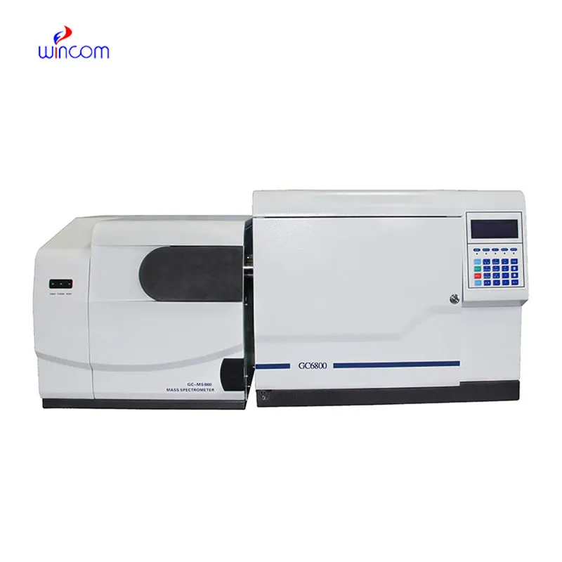
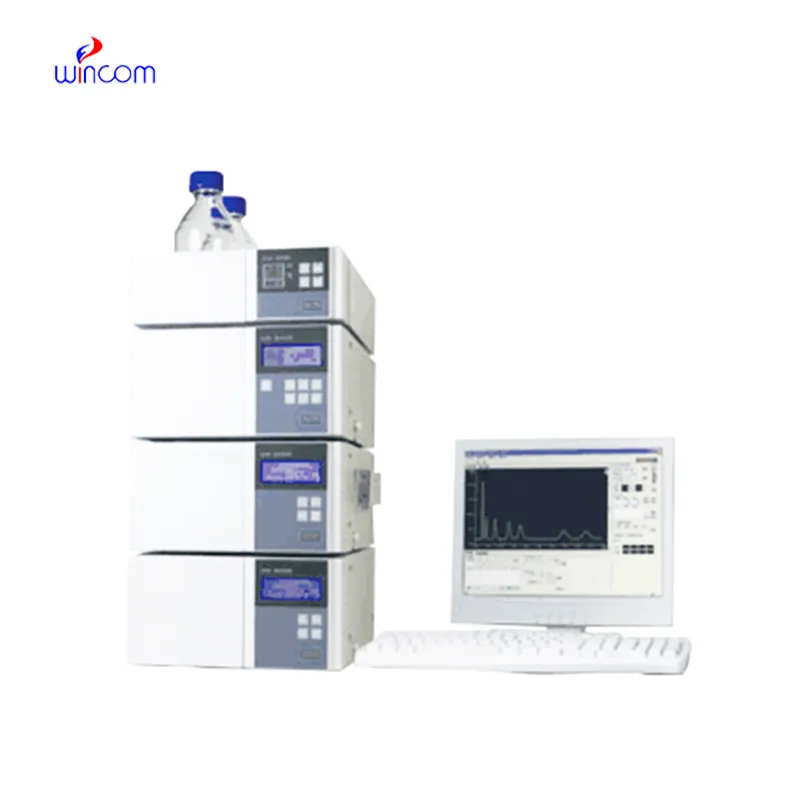
liquid phase chromatography hangs the hospital laboratory in the sense of getting quick and reproducible results for patient sample analysis. Its use is widespread to separate small molecules, hormones, and therapeutic drugs with pinpoint accuracy. Lab staff apply liquid phase chromatography in discovering biomarkers, doing pharmacokinetic studies, and metabolite profiling. Its flexibility makes it suitable for clinical applications with different requirements like research, routine diagnostics, and patient care. So, when hospitals include liquid phase chromatography into their laboratory processes, they get not only the speed but also the dependable analytical performance over various departments.

liquid phase chromatography are utilized by clinical laboratories for hormone and endocrine-related biomarker detection. It delivers trustworthy information for the diagnosis of endocrine diseases by correctly separating substances like cortisol, thyroid hormones, or insulin. Techs in a laboratory rely on liquid phase chromatography to give accurate and repeatable results, thereby helping doctors in individual treatment plan.
The instruments for liquid phase chromatography of the future will be equipped with separation methods in multiple dimensions and fully automated sample preparation. The detection of trace amounts of metabolites, drugs, and biomarkers will be so accurate that hospitals and clinical laboratories will be the first to reap the benefits. The applications of liquid phase chromatography in the future will greatly help in complex diagnostics, research studies, and laboratory efficiency.
Regular system checks, cleaning of detector flow cells, and changing consumable parts whenever necessary are some of the actions that the laboratory staff should take in order to keep the liquid phase chromatography working efficiently. Observing pump performance, taking care of solvent contamination, and storing columns correctly prolong the life of the instrument. Good maintenance assures reproducibility, cuts down on time without access to equipment, and promotes high-quality analysis in hospitals and clinical labs.
Therapeutic drug monitoring relies heavily on liquid phase chromatography in hospital settings. It determines the concentration of drugs in the body to guarantee efficiency and security. The laboratory staff uses it for the examination of blood, serum, or urine samples, and signifies small molecular compounds with high accuracy. By yielding consistent outcomes, liquid phase chromatography services the medics in changing the amounts and preventing side effects. Its use goes to hormone level testing, metabolite analysis, and pharmacokinetics research. With quick processing and accurate information, liquid phase chromatography is a part of the hospital patient care, making evidence-based treatment decisions possible and enhancing clinical outcomes in different departments.
Q: What types of HPLC columns are available? A: Reversed-phase, normal-phase, ion-exchange, and size-exclusion columns are the main types of columns used according to the nature of the analytes. Q: Can multiple samples be analyzed simultaneously? A: Yes, in high-throughput systems, automated sample injection and sequential analysis are among the techniques to achieve this. Q: How does temperature affect HPLC performance? A: Temperature changes can cause variations in separation efficiency and retention times; however, the majority of labs make use of precise temperature control. Q: Can HPLC be integrated with data software? A: Sure, it can be linked with laboratory software for data collection, processing, and reporting. Q: What types of laboratories use HPLC? A: HPLC is employed by hospitals, pharmaceuticals, biochemistry research, and environmental testing labs.
This ultrasound scanner has truly improved our workflow. The image resolution and portability make it a great addition to our clinic.
I’ve used several microscopes before, but this one stands out for its sturdy design and smooth magnification control.
To protect the privacy of our buyers, only public service email domains like Gmail, Yahoo, and MSN will be displayed. Additionally, only a limited portion of the inquiry content will be shown.
Could you please provide more information about your microscope range? I’d like to know the magnif...
Could you share the specifications and price for your hospital bed models? We’re looking for adjus...
E-mail: [email protected]
Tel: +86-731-84176622
+86-731-84136655
Address: Rm.1507,Xinsancheng Plaza. No.58, Renmin Road(E),Changsha,Hunan,China

 af
af
 es
es
 ar
ar
 tr
tr
 sw
sw
 pt
pt
 th
th
 ur
ur
 bn
bn
 ne
ne
 vi
vi
 km
km
 lo
lo
 de
de
 ru
ru
 fi
fi
 nl
nl
 fa
fa
 fr
fr
 ko
ko