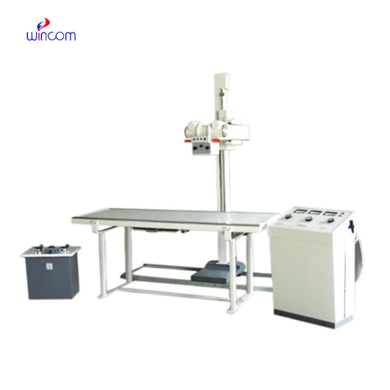
The ray scanner x comes equipped with advanced digital detectors that transform X-ray energy into high definition images of incredible detail. The system's design makes it easy to use and facilitates quick image capturing. The ray scanner x system can be connected effortlessly to hospital information systems that enable the secure transfer of data. The system's robust design provides support for long-term use within healthcare settings.

The ray scanner x is commonly used in medical imaging to examine skeletal trauma, lung disease, and dental anatomy. The ray scanner x assists physicians in diagnosis of fractures, infection, and degenerative disease. The ray scanner x is also used in orthopedic surgery intraoperatively. In emergency medicine, it provides rapid diagnostic information that allows clinicians to assess trauma and internal injury rapidly.

In the years to come, the ray scanner x shall be at the forefront of predictive and personalized medicine. Smart imaging platforms shall look into patient history in conjunction with real-time scans in an attempt to predict upcoming health problems. The ray scanner x shall therefore turn into an anticipatory diagnostic system instead of a reactive one.

The ray scanner x needs regular maintenance to function at its best. Technicians need to regularly inspect exposure controls, cooling systems, and image sensors. The ray scanner x has to be run within prescribed usage boundaries, and annual recalibration needs to be planned to maintain radiation accuracy as well as uniform imaging quality.
In today's healthcare system, the ray scanner x continues to be an integral part of diagnostic imaging. The ray scanner x provides precise visual data that helps in disease detection and assessment of an injury. The ray scanner x has digital sensors and the capability to improve images. The ray scanner x helps in quick and effective medical imaging.
Q: What makes an x-ray machine different from a CT scanner? A: An x-ray machine captures a single 2D image, while a CT scanner takes multiple x-rays from different angles to create 3D cross-sectional views. Q: How is image quality measured in an x-ray machine? A: Image quality depends on factors like contrast, resolution, and exposure settings, which are adjusted based on the target area being examined. Q: What power supply does an x-ray machine require? A: Most x-ray machines operate on high-voltage power systems, typically between 40 to 150 kilovolts, depending on their intended use. Q: Can x-ray machines be used for dental imaging? A: Yes, specialized dental x-ray machines provide detailed images of teeth, jaws, and surrounding structures to support oral health assessments. Q: How does digital imaging improve x-ray efficiency? A: Digital systems allow instant image preview, faster diagnosis, and reduced need for retakes, improving workflow efficiency in clinical environments.
I’ve used several microscopes before, but this one stands out for its sturdy design and smooth magnification control.
This ultrasound scanner has truly improved our workflow. The image resolution and portability make it a great addition to our clinic.
To protect the privacy of our buyers, only public service email domains like Gmail, Yahoo, and MSN will be displayed. Additionally, only a limited portion of the inquiry content will be shown.
We are planning to upgrade our imaging department and would like more information on your mri machin...
Hello, I’m interested in your water bath for laboratory applications. Can you confirm the temperat...
E-mail: [email protected]
Tel: +86-731-84176622
+86-731-84136655
Address: Rm.1507,Xinsancheng Plaza. No.58, Renmin Road(E),Changsha,Hunan,China

 af
af
 es
es
 ar
ar
 tr
tr
 sw
sw
 pt
pt
 th
th
 ur
ur
 bn
bn
 ne
ne
 vi
vi
 km
km
 lo
lo
 de
de
 ru
ru
 fi
fi
 nl
nl
 fa
fa
 fr
fr
 ko
ko