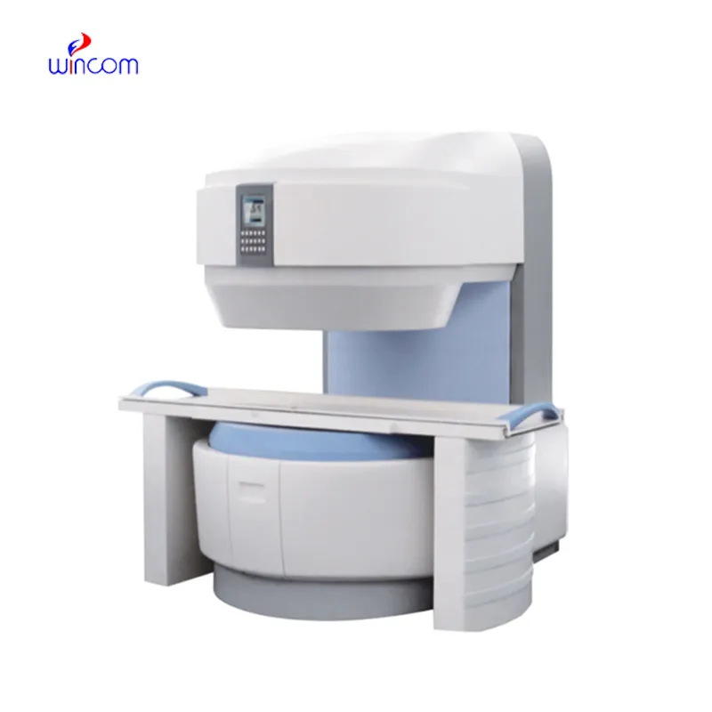
The water path scanner ultrasound, being designed for flexibility, brings the user a variety of imaging modes such as B-mode, M-mode, and color Doppler to use. Because of its small size, it can be easily moved from one place to another and that is why it can be used for examining patients in bed. The water path scanner ultrasound provides good imaging quality, which can be depended upon in cases of routine diagnostics, fieldwork, and emergency medical interventions.

In medical imaging, the water path scanner ultrasound is a trustworthy device for different clinical departments. It provides vascular studies support by observing the state of the arteries and veins; in urology, it has a role in the assessment of the bladder and prostate health. The water path scanner ultrasound also enables emergency medical staff to make quick trauma evaluations, directing their actions precisely and efficiently.

The future of the water path scanner ultrasound may include integration with artificial intelligence for automatic analysis of images. Increased portable functionality and wireless communications may improve remote accessibility in healthcare. The water path scanner ultrasound may include deeper imaging capabilities and rapid data transfer with cloud storage that fosters collaboration on a global medical scale.

Proper care and management of the water path scanner ultrasound needs to be carried out to ensure that it functions well at all times. Cleaning of the probes using a recommended disinfectant helps prevent contamination of the probe and image distortion. Storage of the water path scanner ultrasound in a clean and dry place away from high temperatures helps prolong the life of the equipment.
With advanced transducer technology, the water path scanner ultrasound delivers reliable and high-resolution imaging capability. It allows healthcare providers to see anatomical structures with unmatched clarity and speed. The water path scanner ultrasound enhances workflow efficiency in hospitals, clinics, and mobile medical facilities. With rugged construction and digital connectivity, it makes integration into existing healthcare systems easy.
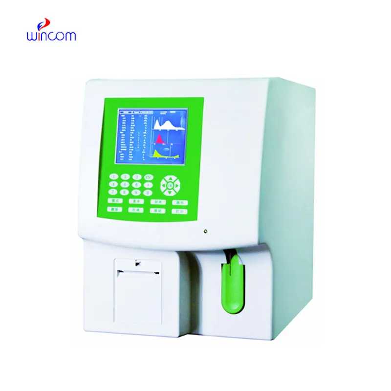
Q: What imaging modes are available on the ultrasound scannert? A: It supports multiple modes such as B-mode, M-mode, and color Doppler for diverse diagnostic applications. Q: How does the ultrasound scannert improve diagnostic accuracy? A: By providing high-resolution images and real-time feedback, it enables more precise medical evaluations. Q: Can the ultrasound scannert be used in field or remote settings? A: Yes, its portable versions are designed for mobility and can be used in clinics, hospitals, or mobile healthcare units. Q: What kind of display does the ultrasound scannert use? A: It typically features a high-definition digital display that enhances image visualization and readability. Q: How is data from the ultrasound scannert managed? A: The device allows secure storage, easy access, and export of imaging data through USB or network connections.
This ultrasound scanner has truly improved our workflow. The image resolution and portability make it a great addition to our clinic.
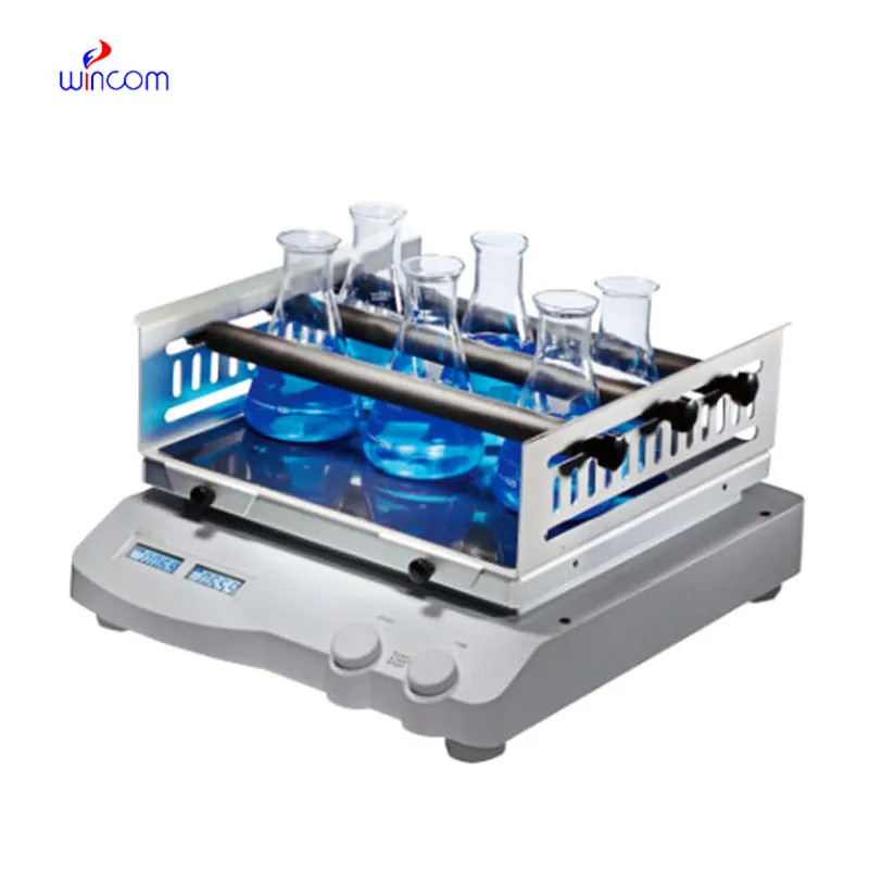
The water bath performs consistently and maintains a stable temperature even during long experiments. It’s reliable and easy to operate.
To protect the privacy of our buyers, only public service email domains like Gmail, Yahoo, and MSN will be displayed. Additionally, only a limited portion of the inquiry content will be shown.
Could you share the specifications and price for your hospital bed models? We’re looking for adjus...
Could you please provide more information about your microscope range? I’d like to know the magnif...
E-mail: [email protected]
Tel: +86-731-84176622
+86-731-84136655
Address: Rm.1507,Xinsancheng Plaza. No.58, Renmin Road(E),Changsha,Hunan,China

 af
af
 es
es
 ar
ar
 tr
tr
 sw
sw
 pt
pt
 th
th
 ur
ur
 bn
bn
 ne
ne
 vi
vi
 km
km
 lo
lo
 de
de
 ru
ru
 fi
fi
 nl
nl
 fa
fa
 fr
fr
 ko
ko