The x ray machine drawing has been designed keeping in mind the needs of modern healthcare. The system comes equipped with capabilities such as real-time imaging adjustments and intelligent exposure. The digital detector provides high uniformity of images, thus supporting accurate interpretations. The x ray machine drawing comes with connectivity solutions that ensure smooth integration with digital radiology networks.

In veterinary practice, the x ray machine drawing analyzes bone fractures, joint diseases, and internal ailments in animals. It provides a transparent image of bone and organ structure, allowing veterinarians to observe health condition precisely. The x ray machine drawing is extremely important in animal health care and research.

Future versions of the x ray machine drawing will combine energy-efficient technology with high-resolution imaging. Predictive analytics integration will enable early disease detection and personalized screening. Global telemedicine networks will also be enabled by the x ray machine drawing, extending access to diagnosis in underserved populations.

The x ray machine drawing require care of the environment and technical inspection. The equipment room needs to be dry, clean, and ventilated well. The x ray machine drawing need to be calibrated regularly, and any unusual sound or display anomaly needs to be reported to technicians at once for evaluation.
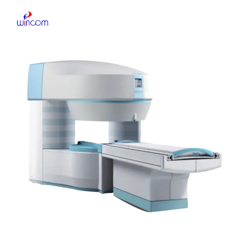
The x ray machine drawing works by providing X-rays that penetrate the body to generate contrast images. The device has been extensively used for screening, diagnosing, and treatment. The x ray machine drawing increases efficiency in healthcare by offering quick images for immediate analysis by a medical practitioner.
Q: What makes an x-ray machine different from a CT scanner? A: An x-ray machine captures a single 2D image, while a CT scanner takes multiple x-rays from different angles to create 3D cross-sectional views. Q: How is image quality measured in an x-ray machine? A: Image quality depends on factors like contrast, resolution, and exposure settings, which are adjusted based on the target area being examined. Q: What power supply does an x-ray machine require? A: Most x-ray machines operate on high-voltage power systems, typically between 40 to 150 kilovolts, depending on their intended use. Q: Can x-ray machines be used for dental imaging? A: Yes, specialized dental x-ray machines provide detailed images of teeth, jaws, and surrounding structures to support oral health assessments. Q: How does digital imaging improve x-ray efficiency? A: Digital systems allow instant image preview, faster diagnosis, and reduced need for retakes, improving workflow efficiency in clinical environments.
The hospital bed is well-designed and very practical. Patients find it comfortable, and nurses appreciate how simple it is to operate.
The microscope delivers incredibly sharp images and precise focusing. It’s perfect for both professional lab work and educational use.
To protect the privacy of our buyers, only public service email domains like Gmail, Yahoo, and MSN will be displayed. Additionally, only a limited portion of the inquiry content will be shown.
I’m looking to purchase several microscopes for a research lab. Please let me know the price list ...
Could you please provide more information about your microscope range? I’d like to know the magnif...
E-mail: [email protected]
Tel: +86-731-84176622
+86-731-84136655
Address: Rm.1507,Xinsancheng Plaza. No.58, Renmin Road(E),Changsha,Hunan,China

 af
af
 es
es
 ar
ar
 tr
tr
 sw
sw
 pt
pt
 th
th
 ur
ur
 bn
bn
 ne
ne
 vi
vi
 km
km
 lo
lo
 de
de
 ru
ru
 fi
fi
 nl
nl
 fa
fa
 fr
fr
 ko
ko